Nailfold Capillaroscopy Research in Raynaud’s
In spring last year SRUK awarded its first research grants, following on from the work of the Raynaud’s and Scleroderma Association and Scleroderma Society, which had been supporting research into both conditions in this way since the 1980s.
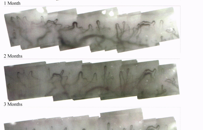
At Hope Hospital, Professor Ariane Herrick and her team have been advancing the use of computerised nailfold video capillaroscopy to monitor changes in capillaries over time in people with primary and secondary Raynaud's phenomenon. Several awards have been made relating to this work covering the period from the early 2000s to 2016.
Software developed by Professor Herrick's team through an RSA grant has enabled capillaries seen under the microscope to be visualised at very high resolution so the state of the same few capillaries can be tracked. The software has been enhanced to measure capillary width, density, shape and form, which assists with disease classification. Studying the same capillaries at each visit means that changes can be identified and monitored. The software is also able to join together high-resolution images so the whole nailfold can be seen and tracked over time.
This tool means that how well treatments work can be seen. The absorption of drugs across the skin can be monitored using vasodilation (the relaxation) of capillaries as a measure of response. It is hoped that the development of a robust scoring system to measure these changes will enable nailfold capillaroscopy to reduce the number of people needed for trials. This will enable the scleroderma research community to evaluate a large number of drugs over the next 5 years.
Who led the research: Professor Ariane Herrick, Hope Hospital and University of Manchester, Manchester
Our Funding: £33,150 (2005-8), £69,799 (2006-9), £158,654 (2011-16)
Tags: scleroderma, Raynaud's, nailfold capillaroscopy
References
- 1.Tian X-P, Zhang. Gastrointestinal complications of systemic sclerosis. World J Gastroenterol 2013;19:7062–8
- 2.Champion HC. The heart in scleroderma. Rehum Dis Clin North Am 2008;34:181-viii